White Paper
Allograft Wedges Biomechanical Impact
Abstract
Introduction
The medial open wedge high tibial osteotomy (MOWHTO) surgery is utilized frequently to correct the varus alignment of the knee caused by medial compartment arthrosis. Although bone graft material has been used to fill the vacuum that occurs following osteotomy, the decision to use bone graft in MOWHTO is still controversial. The aim of this study is to evaluate the impact of the allograft wedge on the biomechanical stability of the medial opening wedge high tibial osteotomy (MOWHTO).
Methods
Two forms of MOWHTO reconstruction were performed: one with a PS SMART Osteotomy HTO plate alone, and the other with a PS SMART Osteotomy HTO plate together with an allograft bone wedge inserted into the osteotomy site (Figure 1). A calibrated servo-hydraulic testing equipment was used to apply a stepwise compression cycle loading, beginning at a maximum load of 800N and increasing by 160N every 20000 cycles. The movement of the proximal tibial fragment was recorded using a 3D motion tracking system (Figure 2). Dynamic stiffness, displacement of the proximal tibial fragment, and rotation have been measured. An independent t-test was used to compare groups under the same loading steps. A paired sample test was used to compare the measurements taken before and after the occurrence of failure within the same group.
Figure 1 | Figure 2 |
|---|---|
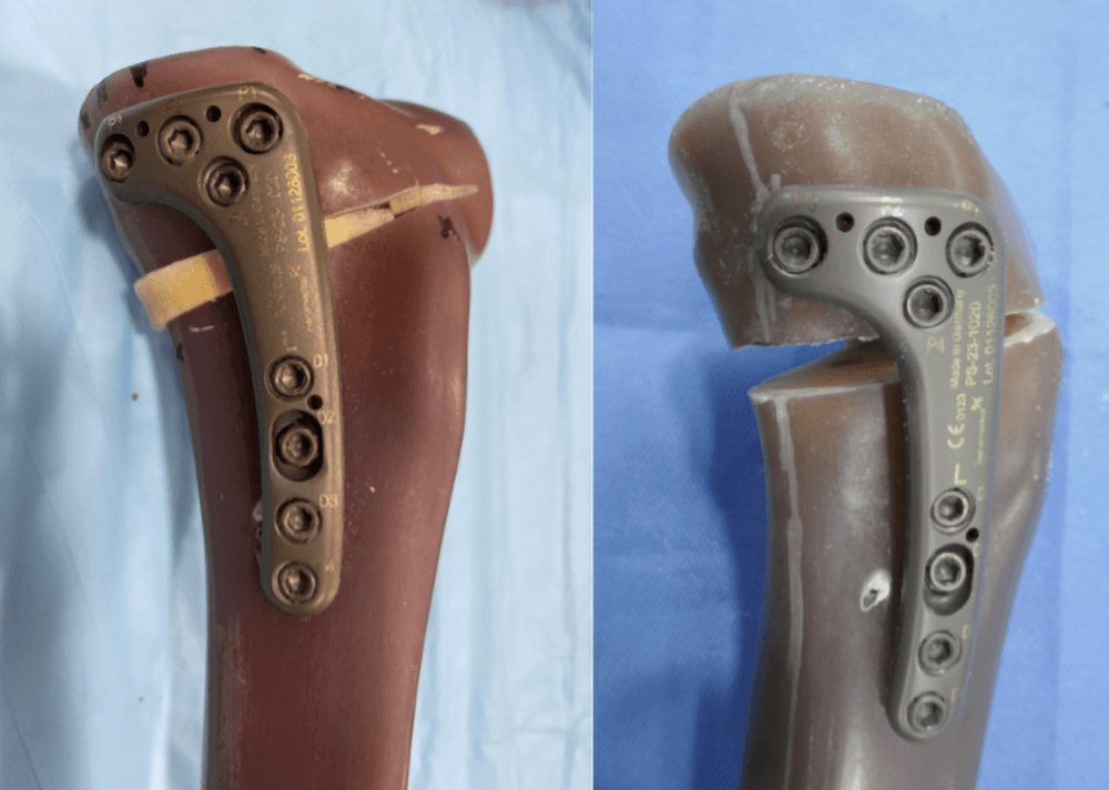 | 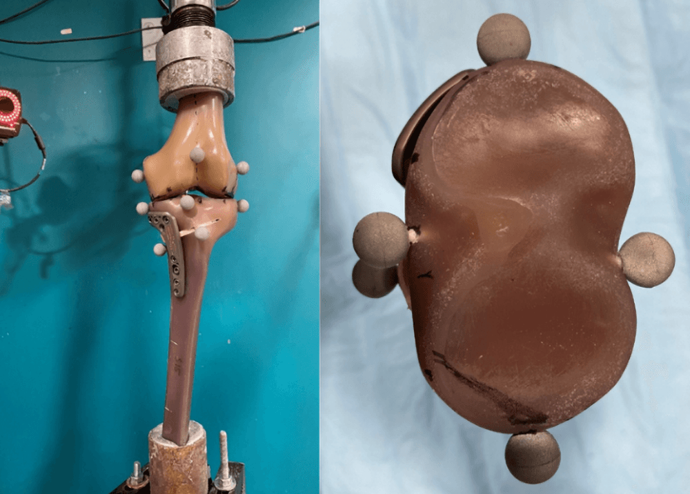 |
Example of sample reconstruction with (left) and without (right) the bone graft filling. | Example of the test setup and marker location on the proximal tibial fragment, left: frontal view of the test setup, right: top view of the tibial plateau, shows the inferred markers were attached to the anterior, posterior, lateral, and medial sites. |
Results
While there was no significant statistical difference between the two groups within the identical loading steps, the bone wedge resulted in a 20% increase in average dynamic stiffness (Figure 3). The proximal tibial fragment displacement of the grafted sample decreased by 17% in the center, 22% medially, and 32% posteriorly (Figure 4). Additionally, the change in the posterior tibial slope angle decreased by 36%. Both groups maintained the proximal tibial fragment valgus angle. The bone graft also reduced the risk of lateral cortex collapse. No significant difference was observed in the average failure load (2360N) and failure cycle (195K) count between the two groups.
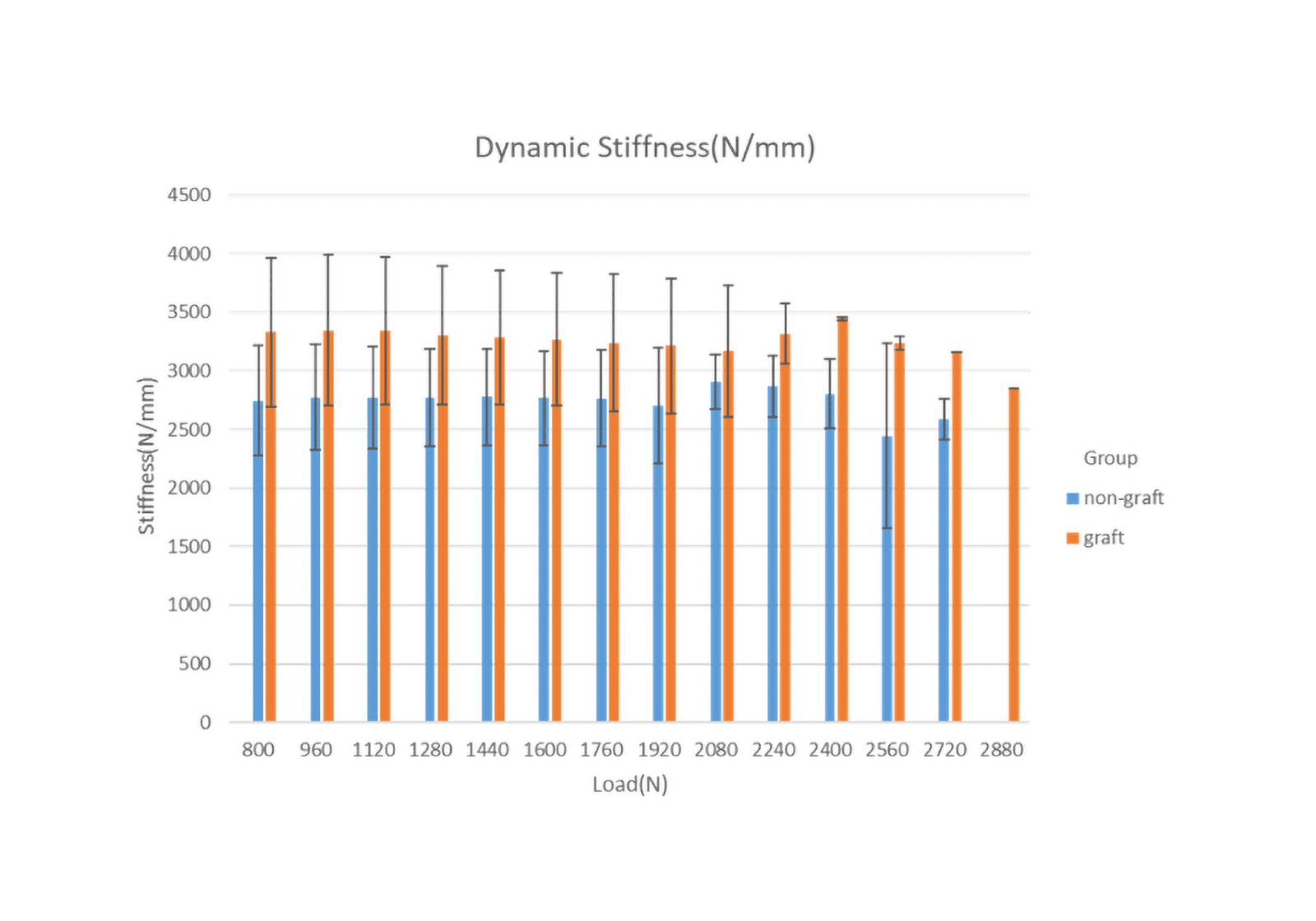
Figure 3 Comparison between non-graft and graft sample dynamic stiffness results under each loading step, error bar indicates +1 standard deviation
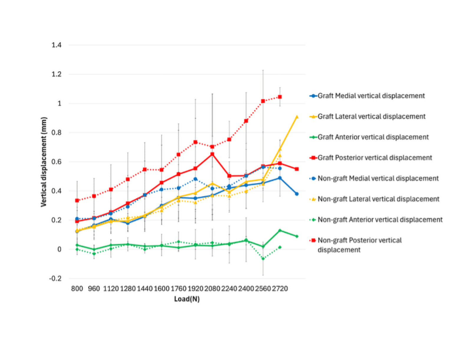
Figure 4. Comparison between non-graft and graft sample medial, lateral, anterior, and posterior vertical displacement results under each loading step, error bar indicates +1 standard deviation.
Conclusion
The findings of the current study provide evidence that the use of bone graft material at the osteotomy site can improve the initial axial and rotational stability of the medial opening wedge high tibial osteotomy (MOWHTO).
Original Research
Post-operative knee adduction moment is associated with patient-reported outcome improvements after medial opening wedge high tibial osteotomy
Original Research
Quantifying Multiplanar Alignment Errors From Patient‐Specific Guide Malpositioning in Medial Opening Wedge High Tibial Osteotomy
Original Research
A newly developed patient-specific instrumentation system achieves consistent alignment accuracy in varising knee osteotomy
Original Research
Accurate and reproducible coronal and sagittal alignment correction using patient-specific instrumentation in medial opening wedge high tibial osteotomy
Original Research
A validated method accounting for joint line convergence angle reduces planning errors in medial opening wedge high tibial osteotomy
Original Research
Accurate correction with a novel patient-specific instrument for medial opening wedge high tibial osteotomy
White Paper
Biologics That Support Bone Healing